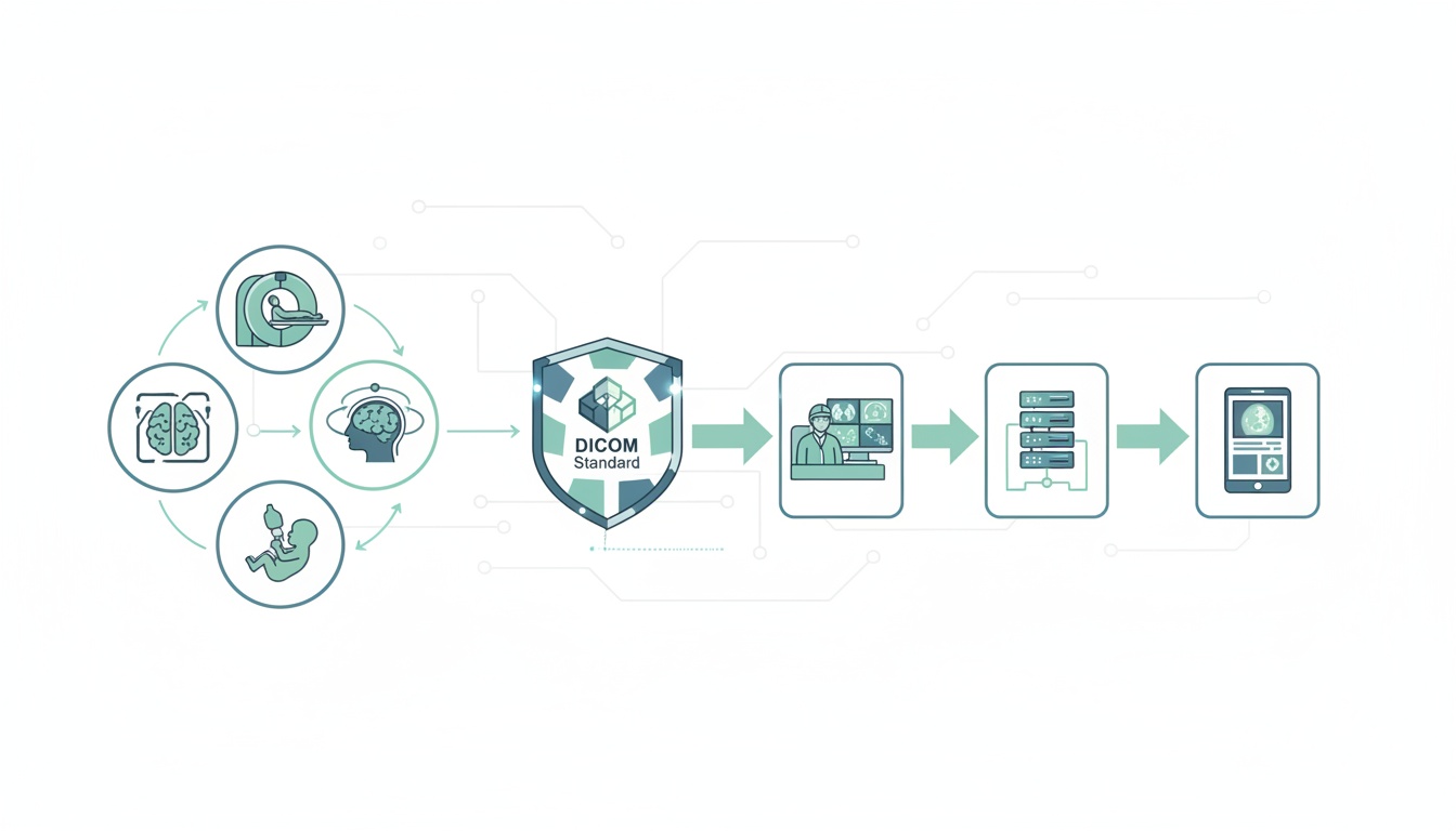
As technology finds its way into every aspect of medicine, great advances have been made in the field of radiology. Radiology once depended on simple two-dimensional images that needed to be manually developed and fixed prior to viewing. Now, almost all forms of medical imaging have become digitalized and the spectrum of radiology includes not just digital radiographs but also CT scans, MRIs, ultrasound, and nuclear imaging. To maintain standards as well as uniformity across the varied types of medical imaging modalities, the concept of DICOM was introduced.
As a medical student or even a full-fledged radiologist, the prospect of dealing with so much technology can seem pretty daunting. This article will explain simply how you can handle DICOM files and exploit their full potential.
DICOM stands for Digital Imaging and Communications in Medicine. It is a standard, internationally accepted format to view, store, retrieve and share medical images. DICOM conforms to set protocols to maintain accuracy of information relayed through medical images.
As a student or practicing radiologist, all medical images that you see are likely to be in the DICOM format. DICOM medical imaging data cannot be opened by regular imaging software present on operating systems such as Windows or Mac OS. A special medical DICOM viewer needs to be installed in order to retrieve, view, and access DICOM medical image files. Therefore, it is important to know how to use this format, what are some relevant applications, and how to access the information and features contained within.
Any DICOM medical image consists of two parts—a header and the actual image itself. The header consists of data that describes the image, the most important being patient data. This includes the patient’s demographic information such as the patient’s name, age, gender, and date of birth. The header may also give information on image characteristics such as acquisition parameters, pixel intensity, matrix size, and dimensions of the image. When a file explorer is opened to view DICOM medical imaging data, the header can give patient and image information. The header is usually coded to the image so that the patient to whom the image belongs can easily be identified. However, the header may sometimes be lost if the DICOM file is exported to other formats, such as JPEG. Sometimes, you may want to intentionally lose the header data, usually for the purpose of anonymization in research cases. This can only be done using specific software functions.
This depends on the purpose for which you want the files. If you are a patient and have had scans taken, you would probably receive a CD or DVD with images on it. If you are a medical student and want to view DICOM images for learning and study, you can download such images from online resources. If you are a radiologist and need to access files in order to interpret them and identify diagnoses, you will need to use a PACS server.
CDs/DVDs: Generally speaking, when medical imaging is done, the patient usually gets a copy of the image files on a CD or DVD. On such CDs, there is usually a DICOM medical imaging viewer included that can help you view the images. Some CDs may not have an actual application, but may provide you with an internet link to download a suitable medical DICOM viewer.
Online resources: If you are a student and are looking for medical images to learn from, there are several online resources that can help you. Some good ones include the Dicom library, Osirix Image library, and the Cancer Imaging Archives.
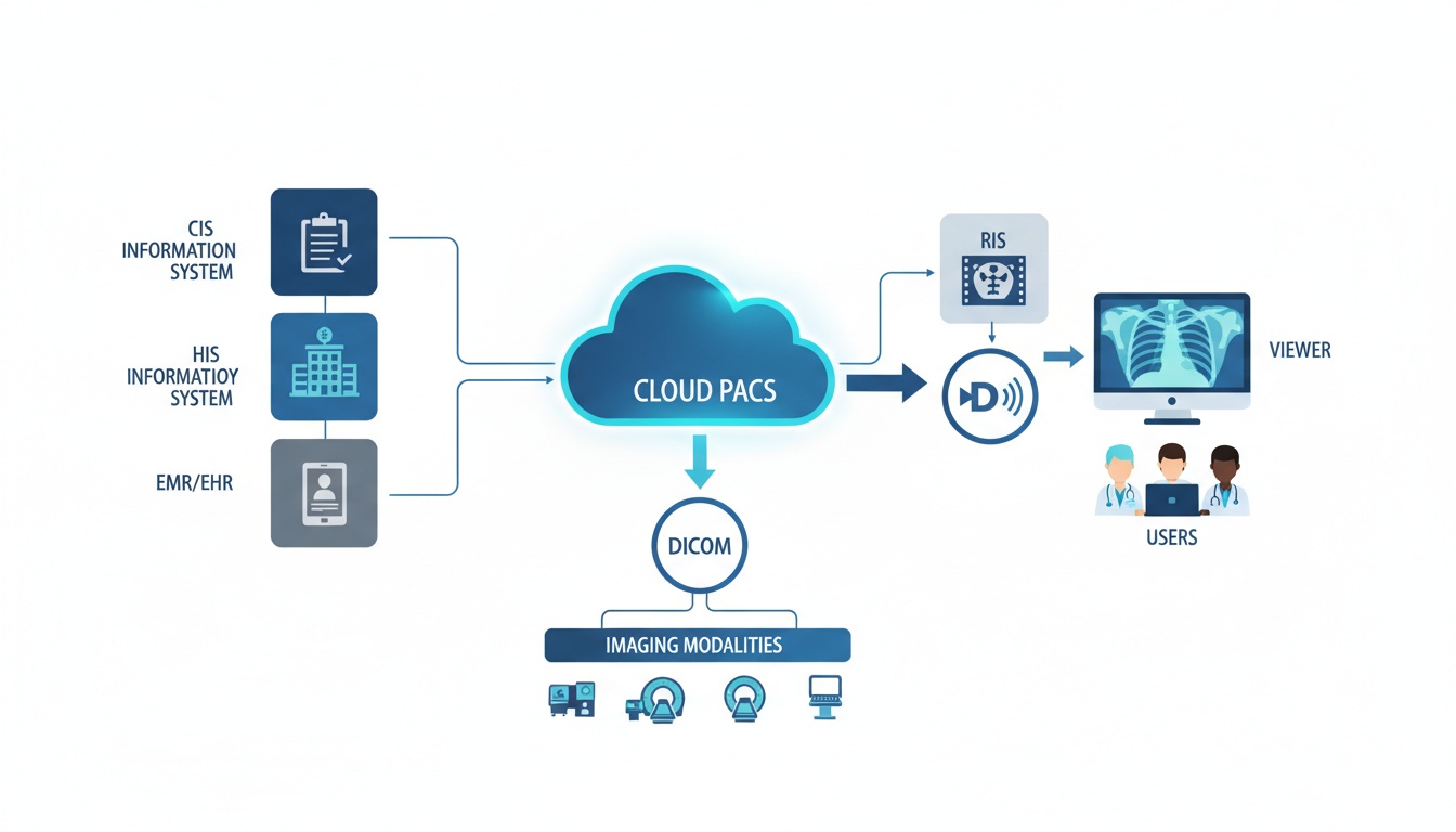
PACS server: PACS stands for Picture Archiving and Communications System. This is basically a database in which all medical images are stored. A PACS server needs to have ample storage, as DICOM files, being of high quality, tend to occupy a lot of space. Each hospital usually has its own PACS server. Any medical image that is taken within a hospital is automatically stored within that PACS server.
For a radiologist to access DICOM medical image files for the purpose of diagnosis and interpretation, a DICOM workstation must be available. This is usually a software application that is capable of complete integration with the PACS server: the application must be able to access and retrieve DICOM images from the PACS server as well as allow viewing and editing and then storing of edited images back again to the PACS server.
|
Cloud PACS and Online DICOM ViewerUpload DICOM images and clinical documents to PostDICOM servers. Store, view, collaborate, and share your medical imaging files. |
Viewing DICOM medical images requires special software. There are basically two kinds of software for viewing DICOM medical imaging data—proprietary software and third party software. Proprietary software comes along with the hardware for medical imaging and is usually created by the same manufacturer. Once the CT or MRI scan is acquired by the machine, the images can be viewed at the same workstation through the DICOM medical imaging viewer. Proprietary software allows users to dynamically view sequential images and also allows reconstruction of these images. One major drawback is that these image files can only be viewed in the same location as the hardware. The images can be transferred to portable storage devices or to other computers on the network only by compressing the images (described in the section below). However, once images are exported, the ability to view and edit the original image is usually lost.
Third party software for DICOM image viewing is becoming increasingly common. These are standalone applications that can open DICOM files from any source—the internet, a CD or DVD, or a PACS server. The market today is flooded with DICOM viewing applications, with both free and paid options available. Each DICOM medical image viewer has its own set of features and you can choose among them depending on your specific requirements.
Today’s DICOM medical imaging viewers can do more than just help you view DICOM images. Some applications are sophisticated enough to improve the image quality as well as generate additional data from the acquired images which can aid in diagnosis. Some of the functions that you can perform, in addition to simply viewing the image, include
Enhancing the image quality: You can increase or decrease the brightness of the image. By changing the contrast, you can distinguish better between radiodense and radiolucent areas of an image. You can also zoom in on the area of interest. A special method of improving image quality in the area of interest involves maximum and minimum intensity projections. This helps isolate areas that have absorbed maximum or minimum radiation, distinguishing them from surrounding areas.
Reconstructing images: The initial DICOM data set contains a series of two-dimensional images, taken in all three planes—axial, coronal, and sagittal. These images can be reconstructed to give a three-dimensional view of the anatomical area. This is called 3D rendering. Another technique, called Multiplanar Reconstruction (MPR), involves making fresh slices from the 3D reconstructed images. This allows the radiologist to view different anatomical levels or angulations from the slices that were originally acquired.
Making measurements: Some medical DICOM viewers allow you to make linear or even volumetric measurements of anatomical structures. This can be useful in planning treatment and assessing the efficacy of treatment. For instance, in cases of traumatic injury to the bony orbit, volumetric analysis and comparison of the injured orbit with the unaffected one can aid in planning orbital reconstruction.
Comparing and combining medical images: DICOM medical applications allow the radiologist to compare two different images at the same time. This is useful when one wants to assess the progress of disease over time or the efficacy of treatment. Two different medical imaging modalities can also be combined using certain DICOM applications. For instance, combining PET and CT images can ensure that areas of high metabolic activity (located using PET) are mapped to specific anatomical sites (using CT scan). This allows the physician to extract the advantages of both types of imaging modalities at the same time.
The DICOM medical file of a single patient consists of multiple images, all of which are of high resolution. Therefore, the file size can be quite large (for instance, a single CT scan can run up to 35 MB). These files therefore need to be compressed before they can be shared and transferred.
There are two ways in which files can be compressed—lossless and lossy. In lossless compression, although the file itself is compressed, there is no loss of information. Therefore, the original file can be recovered at any time. However, this kind of compression requires a lot of processing and so lossless files are slow to open and slow to save. One cannot achieve a substantial amount of compression with this method. On the other hand, lossy compression allows reduction in file size by removing actual data. Usually, only redundant data is removed. Sometimes however, if excessive compression is done, the image quality can be adversely affected. With this method, files can be compressed to much smaller sizes than the original file.
The compressed file can be exported to various formats. Some of the commonest image formats include JPEG, TIFF, PNG and GIF formats.
JPEG (Joint Photographic Experts Group): The JPEG format supports both lossless and lossy compression. Standard JPEG uses lossy compression. The operator can specify how much compression is to be applied and hence can control how much data is lost. Another version, JPEG 2000, supports lossless compression. With this application, operators can identify certain areas of the image as ‘regions of interest’. These areas alone will undergo lossless compression, while other parts of the image will undergo lossy compression. JPEG is useful for easily sharing images between computers. It can be used for uploading images on websites and for standard PowerPoint presentations. On the downside, the quality of the images may not be good enough for paper publications.
TIFF (Tagged Image File Format): The TIFF format can also support both lossy and lossless compressions. TIFF delivers images of higher quality and is therefore the preferred format of several journals for publication. However, the file size is larger and this may not be suitable for use in presentations or on the internet.
PNG (Portable Network Graphics format): This supports lossless type of compression. Using PNG, image features such as brightness and transparency can be controlled. One advantage of PNG is that the images can be tagged using metadata, similar to the header of a DICOM file. This versatile format is suitable for publications, presentations, and the internet.
GIF (Graphics Interchange format): This was one of the earliest imaging formats to be introduced and is not much in use today. It has limited features and the compression is inefficient. It supports lossless type of compression and is largely used for websites.
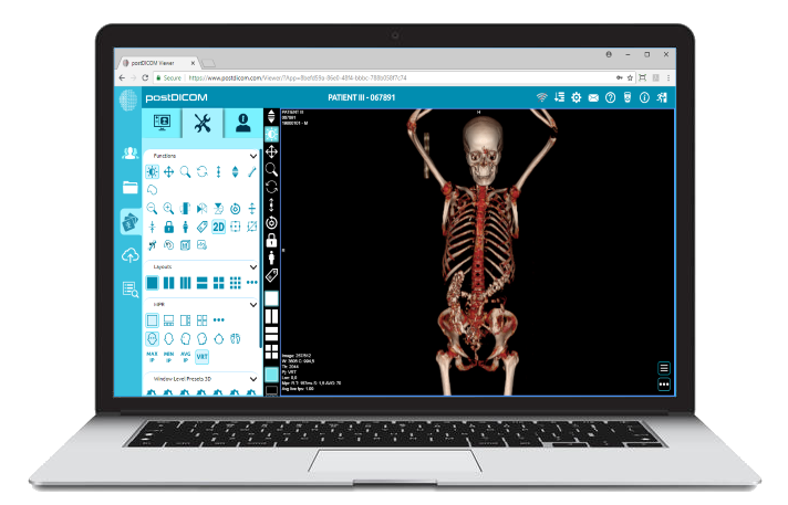
The best way to put your DICOM medical imaging knowledge to use is to go hands-on. The first step is to download a DICOM application that suits your needs, and start using it. There are several free applications that you can use to gain experience. The most popular ones include PostDICOM, Horos, RadiANT, Miele LXIV, and Navegatium.
PostDICOM is a one-size-fits-all application that is just right for the beginner. It is compatible with most commonly used operating systems, including Windows, Mac OS, Linux, and Android platforms. It is quick and has an easy-to-use interface that is great for people who are just learning the basics of handling DICOM images. And at the same time, with advanced features including MPR, MP, MINIP and volume rendering, radiologists can use the application for improved diagnostic capabilities. PostDICOM comes with a unique cloud-based PACS that allows you to store DICOM files online and access them from anywhere, anytime. You can try, PostDICOM medical imaging software free of charge! Through a single accessible account, you can store the images to cloud-based PACS. If you feel that you need more storage space or more user accounts, options to upgrade are available. Try out PostDICOM today!
|
|
Cloud PACS and Online DICOM ViewerUpload DICOM images and clinical documents to PostDICOM servers. Store, view, collaborate, and share your medical imaging files. |